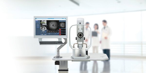
ZEISS CLARUS® 700 Receives NMPA Approval in China, Enhancing Retinal Diagnostics
06 June 2025 | Friday | News

ZEISS Medical Technology announced that the CLARUS® 700 from ZEISS has received National Medical Products Administration (NMPA) approval in China, offering advanced retinal diagnostic capabilities with ultra-widefield, high-resolution images in True Color and unsurpassed quality. The fundus imaging device with Fluorescein Angiography helps eye care specialists in China unlock the full potential of their clinic's retina workflow to support improved patient vision preservation.
"ZEISS CLARUS 700 represents a major step forward in retinal imaging," emphasizes Anuj Kalra, Head of Chronic Disease Management at ZEISS Medical Technology. "By seamlessly integrating ultra-widefield Fluorescein Angiography (FA) imaging with true-color reproduction, this system delivers unprecedented clarity for comprehensive visualization from the macular region to the extreme retinal periphery, enhancing efficiency and supporting precise decision-making within the ZEISS Retina Workflow."
"Integrating ultra-widefield imaging, unsurpassed clarity, and AI-enhanced capture, the CLARUS 700 redefines fundus angiography benchmarks. It will provide unparalleled diagnostic precision for Chinese doctors and unprecedented comfort for their patients," says Maxwell Liu, Head of Sales & Services at ZEISS Medical Technology China.
Ultra-widefield fluorescein angiography serves as a highly valuable examination tool for assessing nonperfused retinal areas. The ZEISS CLARUS 700 HD ultra-widefield fundus imaging camera is an advanced retinal imaging system that provides True Color, high-resolution images. It captures 133°1 in a single image and up to 267° with multiple captures, offering detailed views of the retina. Equipped with both fluorescein angiography and live infrared imaging capabilities, the CLARUS 700 aids in diagnosing and monitoring retinal diseases2. Furthermore, the fundus imaging camera offers innovative technology features including PrecisionFocus for quickly seeing details in the regions of interest, QuickCompare to compare pathology changes observed in past patient visits, and AutoBright so ophthalmologists can spend more time analyzing images and less time adjusting them.
Most Read
- How Does GLP-1 Work?
- Innovations In Magnetic Resonance Imaging Introduced By United Imaging
- Management of Relapsed/Refractory Multiple Myeloma
- 2025 Drug Approvals, Decoded: What Every Biopharma Leader Needs to Know
- BioPharma Manufacturing Resilience: Lessons From Capacity Expansion and Supply Chain Resets from 2025
- APAC Biopharma Review 2025: Innovation, Investment, and Influence on the Global Stage
- Top 25 Biotech Innovations Redefining Health And Planet In 2025
- The New AI Gold Rush: Western Pharma’s Billion-Dollar Bet on Chinese Biotech
- Single-Use Systems Are Rewiring Biopharma Manufacturing
- The State of Biotech and Life Science Jobs in Asia Pacific – 2025
- Asia-Pacific Leads the Charge: Latest Global BioSupplier Technologies of 2025
- Invisible Threats, Visible Risks: How the Nitrosamine Crisis Reshaped Asia’s Pharmaceutical Quality Landscape
Bio Jobs
- Sanofi Turns The Page As Belén Garijo Steps In And Paul Hudson Steps Out
- Global Survey Reveals Nearly 40% of Employees Facing Fertility Challenges Consider Leaving Their Jobs
- BioMed X and AbbVie Begin Global Search for Bold Neuroscience Talent To Decode the Biology of Anhedonia
- Thermo Fisher Expands Bengaluru R&D Centre to Advance Antibody Innovation and Strengthen India’s Life Sciences Ecosystem
- Accord Plasma (Intas Group) Acquires Prothya Biosolutions to Expand Global Plasma Capabilities
- ACG Announces $200 Million Investment to Establish First U.S. Capsule Manufacturing Facility in Atlanta
- AstraZeneca Invests $4.5 Billion to Build Advanced Manufacturing Facility in Virginia, Expanding U.S. Medicine Production
News


